Cytotoxicity - check the quality of being toxic to cells
application for checking the quality
of being toxic to cells
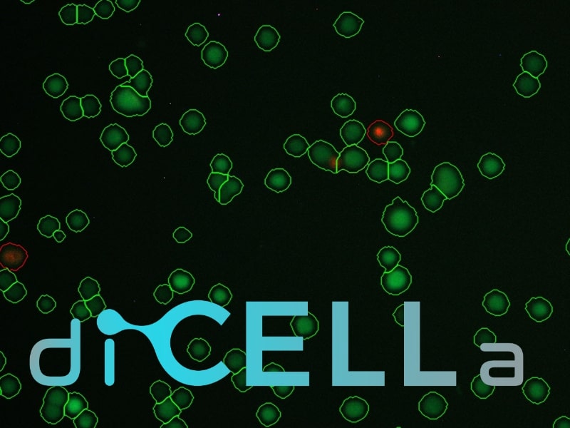
diCELLa Cytotoxicity ASSAY is an application that allows you to quickly analyze results of your experiment. Given only unmodified images our algorithm calculates the live/dead cells ratio and provides you with a number of useful information.
The algorithm:
- is dedicated for fluorescence staining with propidium iodide and AM calcein,
- allows to analyze a wide range of images coming from different imaging devices.
Our solution is fast, automatic, reliable and most of all helps you avoid time-consuming and not always ideal manual analysis.
How to use it?
To use the diCELLa Cytotoxicity ASSAY you need to upload a number of images (max. size 7MB) in any format. The software works on images taken using light microscope and is dedicated for fluorescence staining with propidium iodide and AM calcein.
Results are available to download in txt or pdf format just few seconds after running the experiment in Image Service. They consist of number of live cells, number of dead cells and an output image with marked live cells.
Create an account on our Image Service platform, buy dicellons and exchange them to upload your images.
If you want to use your results in a scientific paper or during a speech at a scientific conference, contact us to get a discount!
Scientific Background
A cytotoxicity assay is incredibly useful in pharmacology and toxicology. In particular, it is used in an oncological research to rate compound toxicity, likewise a tumor cell growth inhibition during drug treatment. Those methods are rapid and inexpensive, however, the evaluation remains non-automatic and tedious for scientists.
It is feasible thanks to the different cells functions, e.g. changes in cell membrane potential, ATP production, co-enzyme production, enzyme activity and nucleotide uptake activity. Experiments in this area are focused on observing how many viable cells are remaining after the drug treatment [1], [2], [3].
One of the most commonly used parameter for this aim is the assessment of cell viability. It is ratio of live cells to all of them expressed as a percentage value [4], [5].
This application allows to make a quick, fully automated evaluation of live/death cells. It is dedicated for fluorescence staining with propidium iodide and AM calcein. The version for a trypan blue staining is under development. Those substances are the most frequently chosen dyes in the cytotoxicity assays [6].
In both staining methods, the state of cells membrane allows to distinguish between alive and dead cells.
How it is working?
In an environment with natural pH, the cell membrane is negatively charged. For this reason, anions are not transmitted through it. If the cell membrane is damaged, the voltage between the outer and the inner side of the cell will decline to 0. For this reason, anionic dyes will be able to delve into inside the cell [7], [8].
In the test with propidium iodide (PI) is being used the calcin AM dye simultaneously. PI marks dead cells by means of staining DNA red in the cell nucleus. On the other hand, AM delves into the cytoplasm and accumulates there in healthy cells, emitting light of green color [9], [10].
The cytoplasm in the test using Trypan blue remains clear for living cells with an undamaged cell membrane. In contrast, the cytoplasm for dead cells is blue-coloured [11].
References:
[1] Aslantürk Ö.: In Vitro Cytotoxicity and Cell Viability Assays: Principles, Advantages, and Disadvantages, 10.5772/intechopen.71923, 2017 December
[2] Longo-Sorbello G. S.A., Saydam G., Banerjee D., Bertino J. R.: Chapter 38 - Cytotoxicity and Cell Growth Assays, Cell Biology (Third Edition), Academic Press, 2006, p. 315-324
[3] Baharith LA, Al-Khouli A, Raab GM.: Cytotoxic assays for screening anticancer agents, Stat Med., 2006 July 15, vol. 25(13): p. 2323-39, PubMed
[4] https://www.uj.edu.pl/documents/2387936/4117775/cw8.pdf, Learning materials from biotechnology, Jagiellonian University
[5] Pegg DE. Viability assays for preserved cells, tissues, and organs, Cryobiology, 1989 June, vol. 26(3), p. 212-31, PubMed
[6] Riccardi C., Nicoletti I.: Analysis of apoptosis by propidium iodide staining and flow cytometry, Nat Protoc. 2006, vol. 1(3), p. 1458-61, PubMed
[7] Traczyk W.: Fizjologia człowieka w zarysie, PZWL, Warszawa 2013, wyd. 8
[8] Suvarna S. K., Layton C., Bancroft J. D. Bancroft's. Theory and Practice of Histological Techniques- 7th Edition, Sheffield 2016 , 173-186
[9] Riccardi Carlo, Nicoletti, Ildo: Analysis of apoptosis by propidium iodide staining and flow cytometry, Nature Protocols, 2006 November
[10] Decherchi P., Cochard P., Gauthier P.: Dual staining assessment of Schwann cell viability within whole peripheral nerves using calcein-AM and ethidium homodimer, Journal of Neuroscience Methods, vol. 71, 1997, p. 205-213
[11] Strober W.: Trypan Blue Exclusion Test of Cell Viability, Curr Protoc Immunol., 2015 November, PubMed